Featured post

Big news: Added 1/8″ inch to wingspan. Phase 1 is complete. Time to move to the next stage.

Here’s the link if there’s issues with the embed: https://youtu.be/Xv_ygWV3dDU?si=c1vrGAMwj4bMUCwb

My initial wingspan was 75 inches. My wingspan measurement is very consistent. I tried torsional loading for like 11 months got no measurable change in wingspan. I even tried manually added some vibration to the exercises no measurable change. I do the exercise above in the youtube video I got 1/16″ in a month. Then I do it for another month about 1/16″ so now my wingspan is about 75 1/8″. Also my thumb has gotten longer based on side to to side comparison with other thumb but there are rotation tricks you can do to fingers appear longer only x-rays are reliable and I have before x-rays but I need to get about 1/4″ to get x-rays because I need sufficient length gain to show up in the x-ray as even with gimp it is hard to detect minor changes.

Essentially, the exercise involves holding the end of a hammer with a pinch grip and then applying vibration to the hand using a machine gun. I do this for at least five minutes a day but often longer up to an hour. The hand does get fatigued doing this though so I have to take breaks. I usually multitask using the free hand.

There is minimal chance that there is measurement error. There is some variance with wingspan measurement but not really when fully stretched. I did torsional loading for 11 months trying to stretch for a result. No gain. I do this exercise for 2 months and the measurement starts creeping up. There is minimal chance that this is measurement error. It’s more likely to be something like soft tissue growth rather than measurement error but soft tissue growth will still be a find. But I still need others to reproduce the result. 1/8″ is small but when measuring something like wingspan or height for that matter once you’ve hit the stretching cap 1/8″ may as well be a mile. Once you’ve perfected stretching your wingspan out the only to increase the measurement is via growth.

The principle behind this exercise is that torsional loading moves fluid within the bone from areas of compression to areas of tension. Vibration makes changes faster so there is more movement than normal. Fluid flow stimulates osteocyte and stem cell activity and this part is not controversial. The controversial part is that it can make the bones longer.

Many anecdotal exercises that increase bone length involve torsion Arm Wrestling, Baseball pitching, Tennis, etc. Tennis also involves vibration as does arm wrestling.

Hiroki Yokota and Ping Zhang favored lateral load over axial loads because logically that is a better way to drive fluid flow within the bone. But torsional loading is superior to lateral loading because what better way to wring water out of a sponge by twisting it.

The exact mechanism by which fluid flow could increase bone length is unknown. But as fluid flow can stimulate osteocytes which can enhance both osteoblast and osteoclast activity which could theoretically remodel the bone to become longer it is possible to see a mechanism. Also the mechanism could involve stem cells as well.

Phase 1 the initial result is the hardest part. Now time to move the next phases:

  1. Get other people to validate the result
  2. Increase my own result so that it can show up on an X-ray
  3. Try to apply the result to other bones.

Here are the keys behind which are needed for the growth to work:

1: The load must be near the bone. If you want to grow the legs for instance squats and deadlifts wouldn’t work because the load is too far from the target bone the legs. The load must be near or on the legs. The reason has to do with direct and indirect loading. If the load is indirect then must of the load is due to muscle pull but if there is a direct loading force than you also get deformations due to the weight itself.

2. The vibration must be near the target bone. The exact location is not important but if the vibration is too far away then the vibrations will dampen before reaching the target bone.

3. The load must be sufficient to put a deforming force on the bone. If it is too light then it won’t drive fluid forces within the bone

Here are some ways to make the exercise more effective:

  1. The load should be asymmetrical. The more the bone is exposed to different regions of tension and compression the more fluid flow is going to flow within the bone
  2. The should be near the epiphysis as the epiphysis is more easily deformable than the diaphysis of the bone.
  3. The loading should be dynamic. The more dynamic the load is the more the regions of tension and compression within the bone will change.

These principles will help in designing exercises for the legs. The torso is challenging because of all the intervertebral discs and the difficulty of applying direct load but it will need to be tackled. It’s also possible to design other exercises for things like the jaw.

But next step is to have other people validate the results. So to validate it I need people to:

Before starting:

  1. Measure wingspan and thumb length and compare hands side by side.
  2. Do the measurement several times to get a feel for potential errors that happen in the measurement.

Then:

  1. Do the exercise in the video for five minutes a day either one hand or both hands
  2. Repeat the measurements done initially weekly.
  3. Report the Result.

Phase 1 is the hardest initial result stage. Now that that is done the result can follow but I need others to validate my result so if you think you an get an accurate measurement and can spare five minutes a day try it out.

Featured post

I was on the Cyborg4Life podcast(again)

Latest:

https://www.youtube.com/live/DayFPZYwNeU?si=2qmpIpe0KcfbV5Z-

I tried not to information dump as much this time but I did last. So I didn’t press him enough on the Kleinburg paper. I also haven’t really explained fluid flow theory so I will have to do a video on that.

Previous:

https://youtube.com/live/7Uu3UO6n8zY?feature=shares

I touched on everything I wanted to touch(obviously not in as much detail) in but I wanted to add that one advantage that an exercise based routine would have over the surgery is that it would provide potentially daily stimulation to bones and could have anti-aging effect as bones are reservoir of stem cells for other tissues.

Myo-inositol to grow taller

This below study indicates that Myo-inositol can increase mandibular condylar cartilage growth but mandibular condylar cartilage can keep growing so we can’t be sure if this applies to long bones but it’s still promising. Myo-inosotol is available for purchase and it’s relatively cheap.

From WebMD “Inositol, also called myo-inositol, D-chiro-inositol, or hexaphosphate (IP6), plays a critical role in the body’s cellular growth. Though it used to be referred to as Vitamin B8, inositol is not actually a vitamin.”

Inositol is already in foods such as beans, bananas, nuts, raisins,and brown rice.

So the question is whether additional supplement is necessary and whether that additional supplementation can result in longitudinal bone growth.

Mandibular Endochondral Growth Is Specifically Augmented by Nutritional Supplementation with Myo-Inositol Even in Rabbits 

“At present, functional orthodontic appliances are used for stimulating mandibular growth in pediatric cases{bite jumping appliance also has potential to work post skeletal maturity}. However, the effectiveness of functional appliances is not always stable in daily practices. A more effective, reliable, and safer therapeutic method for mandibular growth promotion would be helpful for growing mandibular retrognathism patients. As we previously discovered that nutritional supplementation of myo-inositol in growing mice specifically increases mandibular endochondral growth{maybe it could enhance growth of other cartilage} we performed preclinical animal experiments in rabbits in this study. Briefly, six-week-old male Japanese white rabbits were fed with or without myo-inositol supplementation in laboratory chow until 25 weeks old, and 3D image analysis using micro CT data and histological examinations was done. Myo- inositol had no systemic effect, such as femur length, though myo-inositol specifically augmented the mandibular growth{so we have to investigate why that is, perhaps myo-inositol could be combined with other supplements to grow taller}. Myo-inositol increased the thickness of mandibular condylar cartilage. We discovered that the nutritional supplementation of myo-inositol during the growth period specifically augmented mandibular growth without any systemic influence, even in rabbits. Our results suggest the possibility of clinical use of myo-inositol for augmentation of the mandibular growth in growing mandibular retrognathism patients in the future.”

The fact that they think there might be clinical use is promising as it again it is available in foods so it suggests that supraphysiological doses might be helpful.

“A functional appliance is reported to induce mandibular growth by augmentation of endochondral growth in mandibular condylar cartilage and remodeling of the temporomandibular joint”<-this would be amazing if we could get it to happen in other joints and could be the key to growing taller

“nutritional supplementation of myo-inositol in growing mice specifically increases mandibular endochondral growth”<-however mice may not have as diverse a diet as humans

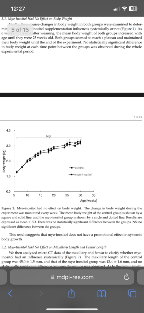
They suggest that mayo-inositol likely did not have systemic effect on growth as there was no change in body weight but if you look at the image you see a slight increase versus control.

However this sentence is not promising “

e myo-inositol group was 90.6 ± 1.1
group was 45.0 ± 1.5 mm, and that of the myo-inositol group was 43.4 ± 1.6 mm, and no mm. There was no statistically significant difference between the groups in femur length.
This result suggests that myo-inositol does not have any promotional effect on bone
statistically significant difference between the groups was observed. As to the femur length,
the control group was 91.3 ± 0.9 mm, and the myo-inositol group was 90.6 ± 1.1 mm.”

However the rabbits(Japanese white rabbits) were given the doses at 6 weeks old and sacrificed at 25 weeks old which is pretty young so it’s possible that growth was slowed but the growing period was elongated. Rabbits stop growing between 5-18months.

Myo inosotol significantly increased mandibular length

“discovered that Pik3cd is specifically strongly expressed in mandibular condylar cartilage”<-maybe we can increase this expression other cartilages to make them grow.

“growth augmentation of the mandible with myo-inositol requires no local injection but just simply needs to supplement the food.”

So the big takeaway here is to make sure you’re getting enough your diet but perhaps we have to find a way to upregulate Pik3cd to grow other joints.

“growth augmentation in the mandible by supplementation of myo- inositol, it was 4.7% induction in our experiments using rabbits. Our previous experiment revealed 8.4% induction in mice”

“inhibition of Pik3cd by a chemical inhibitor almost completely inhibited myo-inositol-mediated augmentation of chondrocyte proliferation in mice”

So next actions for height seekers are definitely to to see if we can increase Pik3cd in other joints to make them more like mandibular condylar cartilage.

New study contributes to evidence that lithium may increase height

One of the mechanisms by which Lithium may increase height is by phosphorylating Gsk 3 Beta. Lithium may also have an impact on the p38 pathway. Lithium also increased proliferation of stem cells.

This study shows directly that Lithium increases growth.

Lithium rescues cultured rat metatarsals from dexamethasone-induced growth failure

“lithium chloride (LiCl) is known to induce cell renewal in various tissues”

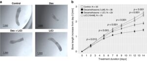
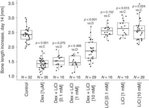
“After 14 days of culture, the length of dexamethasone-treated fetal rat metatarsals increased by 1.4 ± 0.2 mm compared to 2.4 ± 0.3 mm in control bones (p< 0.001). The combination of LiCl and dexamethasone led to bone length increase of 1.9 ± 0.3 mm (p< 0.001 vs. dexamethasone alone). By adding lithium, genes for cell cycle and Wnt/β-catenin, Hedgehog and Notch signaling, were upregulated compared to dexamethasone alone group.”

But they also studied Lithium on its own.

“local side-effects of GCs is the induction of apoptosis in growth plate chondrocytes,5through an increase in the expression of the pro-apoptotic protein Bax”<-Lithium could help reduce cellular apoptosis.

“exogenous GCs have been also shown to impair osteoblast and chondrocyte differentiation via downregulating the Wnt/β-catenin pathway, another key signaling cascade implicated in bone development and local growth plate regulation”<- we know that Lithium affects this pathway.

“Lithium chloride (LiCl) is a known GSK3β inhibitor and the treatment with LiCl increases the proliferation of human mesenchymal stem cells and also rescues from glucocorticoid-induced apoptosis of spontaneously immortalized murine calvarial osteoblasts”

“Bones treated with LiCl alone grew similarly as control (2.55 ± 0.18 mm, 2.64 ± 0.28 mm and 2.59 ± 0.33 mm increase, for 0.1 mM, 1.0 mM and 10 mM LiCl concentration, respectively; versus 2.43 ± 0.25 mm increase for control bones” so you can see that lithium increased growth versus control by about 10% versus the control group! The medium dosage grew the most so Lithium is probably biphasic(there is an optimal equilibrium dose where more has either no effect or is detrimental)

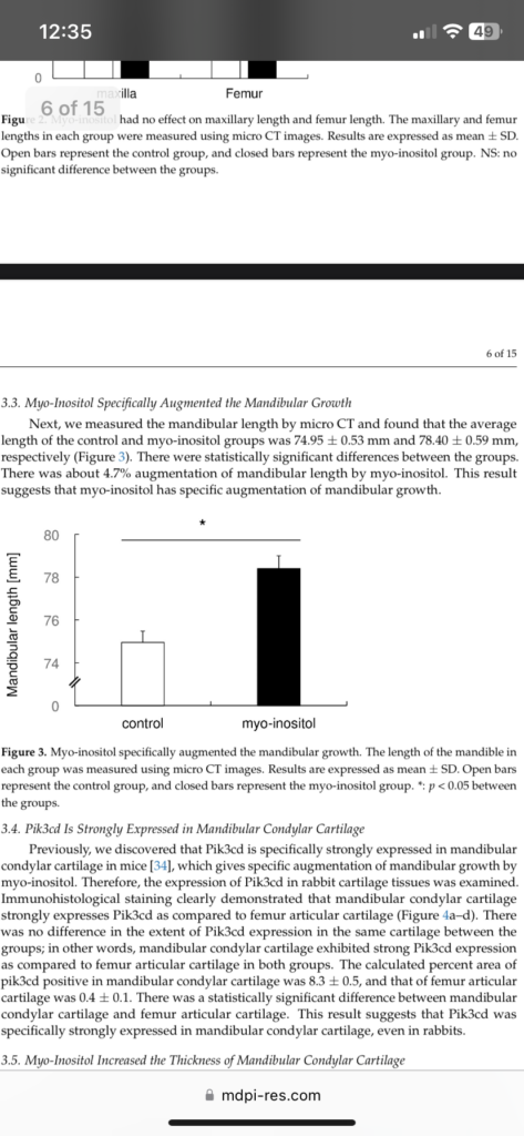
Here’s of what Lithium treated metatarsal looks like via controls:

Here’s another image that shows how significant it is:

“Lithium influenced the expression of 184 genes, of which 98 were up-regulated and 86 were down-regulated”

“a surgical osteoarthritis in vivo model, LiCl provided both in drinking water and intra-articularly was shown to improve the osteoarthritis score and to reduce the severity of cartilage destruction”

“the longitudinal bone growth setting, the results obtained from our proof-of concept experiments are supported by a previous study where lithium carbonate administration increased the width of the proximal tibia growth plate in the domestic fowl. The growth-promoting effect of LiCl was only minor in our “healthy” fetal rat metatarsals, compared to the effect in dexamethasone-impaired metatarsals”

“LiCl upregulates Axin degradation, which leads to β-catenin-induced transcriptional activity, Wnt/β-catenin pathway seems to be a plausible target that could explain the growth-promoting effect of LiCl in the dexamethasone-impaired metatarsals.”

“higher doses of LiCl have clinically been associated with undesired side effects such as tremor, dizziness, nausea, polyuria, weight gain, hypercalcemia or hypothyroidism, which therefore limits the use of high-dose LiCl treatment”<-again Lithiun is biphasic

So looks Lithium could be a height increase supplement and it is available to people!

This paper shows why lateral and torsional loading are so effective in causing bone adaptation

A Wolff in sheep’s clothing: Trabecular bone adaptation in response to changes in joint loading orientation

Most loading to bones is axial but there is always some non-axial loads as the bones have shape thus there is a mix of tensile, compressive, and torsional loading. But loading bones in one axis will tend to produce the same stimuli, whereas doing things like lateral and torsional will induce unique stimuli. This will produce more effective adaptations including possibly growing taller if the loading is sufficient.

“This study tests Wolff’s law of trabecular bone adaptation by examining if induced changes in joint loading orientation cause corresponding adjustments in trabecular orientation. Two groups of sheep were exercised at a trot, 15 min/day for 34 days on an inclined (7°) or level (0°) treadmills. Incline trotting caused the sheep to extend their tarsal joints by 3–4.5° during peak loading (P b 0.01) but has no effect on carpal joint angle (P= 0.984). Additionally, tarsal joint angle in the incline group sheep were maintained more extended throughout the day using elevated platform shoes on their forelimbs. A third “sedentary group” group did not run but wore platform shoes throughout the day. As predicted by Wolff’s law, trabecular orientation in the distal tibia (tarsal joint) were more
obtuse by 2.7 to 4.3° in the incline group compared to the level group;{Can this change in trabecular orientation effect bone length?}
trabecular orientation was not significantly different in the sedentary and level groups. In addition, trabecular orientations in the distal radius (carpal joint) of the sedentary, level and incline groups did not differ between groups, and were aligned almost parallel to the radius long axis, corresponding to the almost straight carpal joint angle at peak loading. Measurements of other trabecular bone parameters revealed additional responses to loading, including significantly higher bone volume fraction (BV/TV), Trabecular num-ber (Tb.N) and trabecular thickness (Tb.Th), lower trabecular spacing (Tb.Sp), and less rod-shaped trabeculae (higher structure model index, SMI) in the exercised than sedentary sheep.
Overall, these results demonstrate that trabecular bone dynamically adjusts and realigns itself in very precise relation to changes in peak loading direction{it’s also possible that mixing up axial, lateral, and torsional loading could cause the trabecular to have to constantly dynamically adjust}, indicating that Wolff’s law is not only accurate but also highly sensitive”

“changes in posture will alter stress distribution within the bone and eventually, if they persist, the trabecular structure will readjust to the new stress trajectories.”

The paper mentions that different locomotive methods induce changes in trabecular orientation.

“As intended, the angle of the tarsal joint was more extended by 3.6° at
the time of peak GRF (midstance) in the incline group”

From the looks of this image the incline group looks like it stands taller even though bone length may be the same.

“a minimum level of loading is necessary to affect trabecular growth is evident from the almost total lack of trabeculae documented in the sagittal
ridges of the distal tibia and radius”<-So there must be sufficient weight used in lateral and torsional loading regimes.

“An additional limitation is that we studied only very young animals whose joints were still growing and remodeling.”

Note how much bigger and longer the epipihysis is in image B versus A but again these are growing animals. They said that trabecular did not really change much in the radius(groups A and B) but the two radius look much bigger.

In contrast the Group D looks smaller than group C which is the tibiae which is what changed.

Studies like Trabecular bone in the calcaneus of runners, were conducted on runners above age 20 so it’s likely still that trabecular bone is responsive to the direction of loading.

The paper Physical activity engendering loads from diverse directions augments the growing skeleton, shows that again loads from diverse directions are important thus perhaps typical axial loading is enough.

“Growing mice housed for three months in cages designed to emphasize
non-linear locomotion (diverse-orientation loading) were found to have enhanced trabecular and cortical bone in the proximal humerus compared to animals housed in cages that accentuated linear locomotion (stereotypic-orientation loading).”

These papers show why we must try to use experiment with lateral and torsional loading methods in attempts to grow taller.

Study shows that mechanical loading can alter the trabecular bone via the growth plate

This study shows that mechanical loading can alter the growth plate and the trabecular bone. This shows it is possible to use mechanical loading to alter height during development and also perhaps spinal height as there is a great deal of cartilage via the discs there.

Unveiling the Trabecular Connection: Exploring Morphological Adaptations in the Growth Plate and Their Response to Mechanical Stimulus

“The growth plate, also known as the epiphyseal plate, is a critical element in the longitudinal growth of long bones during development. This study aims to explore the connection between trabecular patterns and morphological alterations within the growth plate, particularly concerning their impact on shape and mamillary processes or trabecular patterns as well as the formation of ossification bridges{ossification bridges has the potential to reduce growth so studying them has potential}. Our objective is to investigate how the adaptations in the growth plate and the trabecular bone in its vicinity respond to mechanical stimulus, this approach is considered a new methodology to study endochondral growth and bone remodelling”

“Our investigation revealed insights into the development of the growth plate and its capacity to adapt its shape in response to the local mechanical environment. Previously, this environment had been predominantly modeled as a continuum, but our model allowed us to assess the impact of localized load transmission via the trabecular groups. Furthermore, our research demonstrated that the morphological changes within the growth plate in addition to bone adaptation in its vicinity, serve as an adaptive mechanism to withstand shear stress{so we would want to optimally induce shear stress, I believe that torsional loading is the best way to induce fluid shear}, contributing to increased bone density in specific regions”

“at birth the growth plates have a flat and smooth topology in humans, as age increases a wavy pattern is seen, this change is due to mechanical stresses”

“The thickness in the growth plate remains constant during childhood thanks to an equilibrium between chondrocyte proliferation and apoptosis. In puberty, when this balance is perturbed, the process of growth plate fusion starts, forming ossification bridges in different locations until the entire plate has been ossified, leaving behind a bone remnant, formally known as epiphyseal scar”

“animals, such as mice and rats, which are widely used as in-vivo models for endochondral ossification studies, do not entirely close their growth plates during maturation”<-but their growth plates do seem to be become senescent which indicates that there may be epigenetic factors that limit growth plates from growing indefinitely.

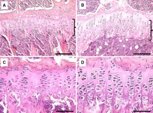
this looks like an example of the histological changes due to lateral synovial joint loading so joint loading definitely alters the growth plate morphology:

“the change in shape in the growth may occur as a consequence of the bone remodeling process in the vicinity of the growth plate, and at the same time trabecular formations adapt”

“A future study case for this model will be a therapy of hemiephysiodesis or guided growth, where this model can be used to quantify the degree of growth modulation along the entire growth plate, or the
change in geometrical descriptors; simply by including a transphyseal screw in the domain.”<-so there is potential for this research to be used for people to grow taller.

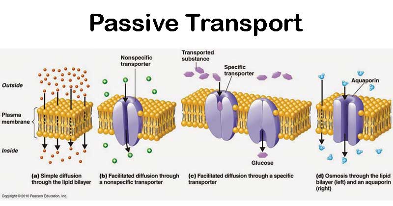
Passive diffusion and it’s potential role in growin taller

If articular cartilage endochondral ossification(and) is to be a viable with which to grow taller then the existing articular cartilage must grow and replenish while the rest ossifies. Vertebral and joint cartilage contributes to height so if there was a way to get them to grow then we could become taller. Since cartilage has poor blood supply it heals and grows by passive diffusion. So our goal would be to maximize passive diffusion.

Passive diffusion is where a molecule from high concentration to low to concentration.

The factors affecting diffusion are :

Temperature: Higher temperatures increase molecular energy, leading to faster movement and enhanced diffusion.

Concentration Gradient: The steeper the gradient (difference in concentration), the faster the diffusion.

Molecular Mass: Smaller molecules diffuse more rapidly than larger ones.

Medium: Different substances diffuse at varying rates through different media.”

So what we can influence is temperature and concentration gradient. Temperature is obvious but for concentration gradient we can increase supplementation and increase fluid flow as the more fluid is flowing the higher concentration is going to be and therefore the more compounds are going to be diffusing into the articular cartilage.

This coincides with anecdotal evidence that movement is vital for healthy cartilage and with the observation that sufficient force in chiropractic treatment is required to induce joint cavitation.

The Effect of Antibody Size and Mechanical Loading on Solute Diffusion Through the Articular Surface of Cartilage

” Therefore, the goal of this study is to investigate how the size of antibody (Ab) variants, as well as application of cyclic mechanical loading{the cyclic nature of the loading as the changing in load rate is important as well as overall load}, affects solute transport within healthy cartilage tissue. Penetration of fluorescently tagged solutes was quantified using confocal microscopy. For all the solutes tested, fluorescence curves were obtained through the articular surface. On average, diffusivities for the solutes of sizes 200 kDa, 150 kDa, 50 kDa, and 25 kDa were 3.3, 3.4, 5.1, and 6.0 μm2/s from 0 to 100 μm from the articular surface. Diffusivities went up to a maximum of 16.5, 18.5, 20.5, and 23.4 μm2/s for the 200 kDa, 150 kDa, 50 kDa, and 25 kDa molecules, respectively, from 225 to 325 μm from the surface. Overall, the effect of loading was very significant, with maximal transport enhancement for each solute ranging from 2.2 to 3.4-fold near 275 μm{so mechanical loading has a lot of potential to benefit passive diffusion and therefore cartilage growth}. Ultimately, solutes of this size do not diffuse uniformly nor are convected uniformly, through the depth of the cartilage tissue. This research potentially holds great clinical significance to discover ways of further optimizing transport into cartilage and leads to effective antibody-based treatments for OA.”

Rodeo may be another exercise that increases longitudinal bone growth

Bareback riding is horseback riding without a saddle.

As seen in the picture below bareback rodeo riding involves asymmetrical loading on one arm and it involves torsional+vibrational loading at the arm.

There is anecdotal and some study evidence that exercises that involve torsional+vibration loading such as tennis and arm wrestling can increase longitudinal bone growth even past epipihyseal fusion. Since rodeo has vibration + torsional loads the question is can it increase longitudinal bone growth as well? In short: the study below suggests that rodeo riding can increase bone size but longitudinal bone length was not measured. The difference in bone size is very large though so it looks to be an effective stimulus. I did send an email to the author of the study though.

Bony hypertrophy of the forearm in bareback rodeo athletes

“Activities that require increased load bearing are known to cause bony hypertrophy. This phenomenon has been documented in the dominant arm of athletes in sports requiring significant utilization of a single limb. The literature addressing this effect in rodeo athletes, however, is minimal. Studies evaluating rodeo athletes are primarily focused on acute injury management rather than chronic symptoms resulting from changes in bone and soft tissue. We designed a study to evaluate bony hypertrophy in athletes without acute injury.

Rodeo bareback riders presented with frequent pain in their grip arm, no radiographic evidence of injury, and clinical signs of peripheral nerve compression. Anteroposterior and lateral X-rays taken for initial evaluation in 17 bareback rodeo athletes were retrospectively reviewed. The diameter of bilateral ulnas was measured at its longitudinal midpoint. Ratio of Ulnar Diameters (grip arm/free arm) and Percentage Diameter Difference were calculated. An independent samples t-test was used to assess differences in diameters of grip and non-grip arms.

The mean ulnar diameter was 18.4 ± 3.5 in the grip arm and 16.6 ± 3.5 in the non-grip arm {this is a pretty big difference}. The mean ratio of grip to free arm ulnar diameter was 1.42 ± 0.21 (range = 1.05–1.92). The mean diameter percent difference measured 42.3% (range = 4.7%–92.0%), and the grip arm was observed to have a greater ulnar diameter compared to the non-grip arm.

There are significant anatomic differences in the grip arm of bareback rodeo athletes compared to the contralateral arm. In cases of persistent pain in the grip arm and no evidence of acute injury, these differences may be relevant to pain symptoms and should be considered as part of the assessment and treatment algorithm.”

This paper cites the paper “Chronic Hypertrophy of the Ulna in the
Professional Rodeo Cowboy”. Here is an image of that paper

The left ulna looks much wider at the epiphysis. Possibly longer.

Going back to the paper. Here’s an x-ray from the paper not the difference in bone width

Here’s an image from the paper comparing the two arms:
Left arm looks longer but we’d have to additional studies to see.

“In this study, we show that bony hypertrophy in the ulna of the grip arm of bareback rodeo riders was 42.3% greater on average when compared to the non-grip arm, which is nearly a 10-fold greater difference (between right and left) in forearm bone thickness than a previous study on tennis athletes.”<-so rodeo training may be more effective than tennis. We’d have to think about this when designing bone lengthening exercise protocols.

“This study is limited by the inclusion of only symptomatic patients. We were unable to account for previous injuries that may have influenced the bone composition of either arm. Furthermore, we are unaware if the athletes participated in other activities requiring increased stress to the grip arm. This study did not include athletes from other sports (e.g. tennis, baseball, or hockey) for direct comparison of the degree of hypertrophy seen in rodeo versus other sports.”<-So there’s selection bias and they can’t control for whether only rodeo was responsible for the effects. The athletes could have done other training that contributed.

Rodeo is an expensive sport so it’s not really something that we can self-test effectively and it’s hard to control the stimulus as it’s really based on what the bull is doing. But bull riding can provide additional evidence that torsional/vibrational loading can increase longitudinal bone growth. In this study, the grip arm does look longer so that is anecdotal evidence but that could be an illusion. This is definitely worth diving into further,