The Biological Imaging Facility is a core microscope imaging facility that specializes in widefield fluorescence, laser scanning confocal, spinning disk confocal, TIRF, and super-resolution microscopy (Lattice SIM, PALM, STORM), as well as traditional plant & animal microtechnique, histology, and cryotomy.
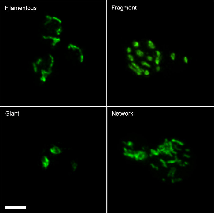
Maximum intensity 3D images stacks of different mitochondrial morphologies in yeast. Mitochondria expressing GFP were imaged using the Zeiss 880 confocal, then the data stacks were deconvolved using SVI’s Huygens and analyzed with Bitplane’s Imaris. All of these instruments and programs are available for use in the Biological Imaging Facility. Scale bar is 2µm
Jun-Ting Johnson Wang of the Brem Lab
Jun-Ting Johnson Wang of the Brem Lab
The Rausser College of Natural Resource’s Biological Imaging Facility functions as an instructional and research laboratory for all aspects of modern light microscopy, including confocal and super-resolution microscopy, image processing and analysis, and most microscopical techniques for developmental and cell biology. Computer image processing and analysis is taught on an individual basis. Microscopy is taught on two levels:
- Individually as needed
- PMB185 Techniques in Light Microscopy.
BIF Training and Use Policy
There are full time staff members who provide training in the correct use of the advanced microscopes in the BIF. We also provide training and consultation in advanced techniques of microscope imaging, image processing and analysis.
- If you wish to be trained, please complete this Google Form, then contact Denise Schichnes. Once you have completed this training form and are trained, you will be authorized to reserve instrument time and enter the BIF.
- If you are already trained feel free to reserve instrument time yourself. Contact us for the Calendar signup credentials.
There are four core laboratories on campus that offer expertise, instruction, and instrumentation in microscopy for research.
- The CNR Biological Imaging Facility (This lab): widefield, confocal, and super-resolution epifluorescence microscopy, live-cell imaging, microtechnique, training in digital image processing and analysis. Located in 381 Koshland Hall
- Molecular Imaging Center: Two-photon, SPIM, confocal microscopy, electrophysiology, live-cell imaging, and FLIM. Located in Weill, Li Ka Shing, and Barker Halls.
- Electron Microscope Laboratory: Transmission and scanning electron microscopy. Located in Barker Hall.
- CIRM/QB3 Shared Stem Cell Facility: Available for the study of stem cells. Located in Stanley Hall
- The Golub Microscope Collection: Web site dedicated to Dr. Orville J. Golub’s collection of antique microscopes from the 17th–20th Centuries. The collection is located in VLSB. Dr. Steve Ruzin is the Curator.